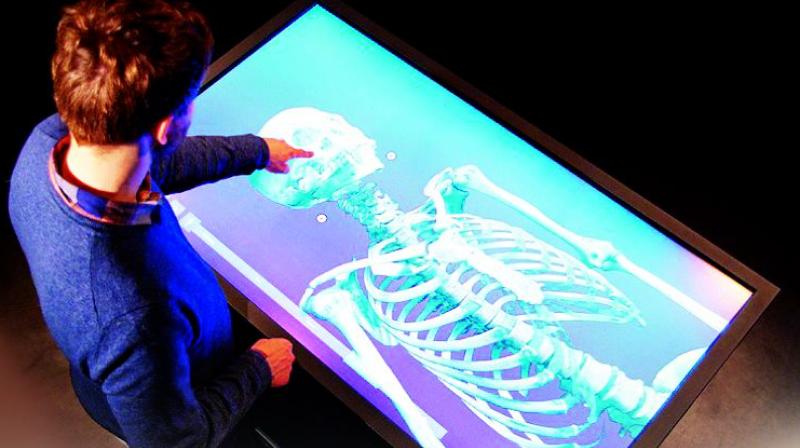
Going Digital: Medicos look for hi-tech autopsy
AIIMS example has colleges hopeful

Hyderabad: Virtual autopsies are being encouraged for teaching purposes in medical colleges, as it gives a clearer picture of fractures, tumours and infections suffered by the body. Virtual autopsy is a hi-tech digital X-ray and minimally-invasive procedure wherein the body of the dead does not have to be slit at various parts, as is generally done. But it still gives a clear understanding of diseases, injuries and even the minute clot, if any.
The introduction of virtual systems at the All India Institute of Medical Sciences has enthused the anatomy department of both private and government medical colleges, and they are looking forward to introduce the new technology in their institutions. However, as of now, they are having a tough time sourcing a body for studies.
Dr C. Mrudula, professor and head of department of anatomy at Apollo Institutes of Medical Sciences and Research, explained, “The three - dimensional approach allows us to visualise even the minutest foreign object in a body. It allows measuring the depth and also gives the exact location. This method is better than the one wherein professors have to see things inside the body with their naked eyes.”
With more than 150 students in a medical class, teaching them anatomy on one body is difficult. Mostly, professors have to create models which will give students a feel of what it’s really like. The virtual system has come as a boon, as it helps understand the procedure on a wider and bigger screen. It can be replayed in case of doubt.
The possibility of re-learning by referring the cases later is easy and feasible. In some cases, the bodies available are highly decomposed or infected with HIV or the like. A senior professor said, “In case of these bodies, cutting them deep is not possible as there’s the fear of cadaver infections. In such cases, virtual autopsy helps.”
The advantages
- It gives a complete and non-destructive
- information from head to toe.
- It reaches the places in the body where traditional methods cannot approach.
- It helps for re-examination if we require a second opinion.
- Clean, bloodless visualisation of the documentation.
- Bodies that are toxic can be easily examined without any contamination
- Acceptance by relatives and religious communities.
- It is time-saving.
- It allows us to identify foreign bodies present.
- X-rays give the edge detail of radio-opaque or metallic objects, one can sort out what the object might be, and CT, because it is three-dimensional, shows you where the object is in the body.
- Not only can you preserve the data but you can also “share” or collaborate on that autopsy in medical education.
The Disadvantages
- High costs.
- All the pathological changes cannot be visualised.
- It’s difficult to determine colour changes of all internal organs.
- Vascular and metabolic changes cannot be seen.
- It requires specialised staff and faculty.

