Complex heart surgeries simplified
Raj turned blue a few days after his birth as great arteries were transposed, resulting in poor oxygenation of blood.

CHENNAI: Raj Kumar, a newborn at a city hospital, was diagnosed with a cardiac disorder often called as a blue baby syndrome that indicated cardiac complications. Raj turned blue a few days after his birth as great arteries were transposed, resulting in poor oxygenation of blood.
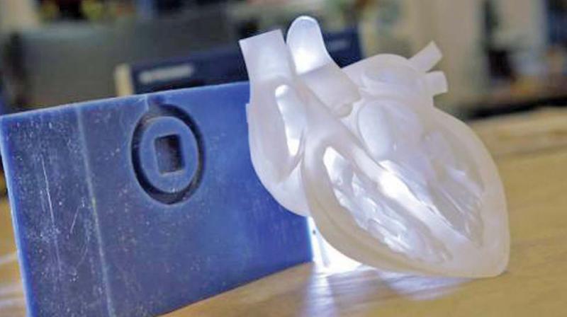
His valves were also found to be defective, 3-dimensional (3D) printing technology came to his rescue and he underwent an open-heart surgery after imaging his heart using the technique. Raj recovered soon and is completely normal after five years of the surgery.
Around 10,000 children born with congenital heart diseases in Tamil Nadu and 90 per cent of child heart patients do not receive timely attention, resulting in premature death or lifelong disability. However, the imaging technique helps to make a 3-dimensional model of the heart, serving as a template for the cardiologists and cardiac-surgeons to perform the surgery.
“Most heart defects need open-heart surgery, even in newborns. The maximum number of deaths from congenital heart defects happen in the first year after birth and a substantial proportion of them die in the first month of life,” head, department of paediatric cardiology, Amrita Institute of Medical Sciences.
When requiring a surgery in paediatric patients, 3D heart models are necessary as it is more complex owing to its size. The most common congenital heart defects in children include holes in the heart or the great vessels, the narrowing of heart valves or vessels, and conditions resulting in the child becoming a “blue baby”.
“In case of congenital heart diseases, after screening the heart using a magnetic resonance imaging, the 3D printer produces an actual image of a similar heart as of the patient. This provides a clear-cut analysis of the complexity of the defects and surgery can be done accordingly,” said Dr Suresh Rao, a senior cardiac surgeon.
3D printing technology also helps in case of transplants in paediatric patients, as the size of the donor's heart might not fit in the chest cavity of the receiver. 3D printing can help to produce a live model of the requirement of the donor after imaging. “3D imaging technique in case of cardiac surgeries can aid to produce patient-specific surgical models that are needed to prevent congestive heart failure due to ventricular dysfunction. Similarly, in cases of primary tumours of heart, imaging and 3D printing can support the planning of the resection and aid in surgical repair,” said Dr S. Balasubramaniam, a cardiologist.
Congenital heart disease Diagnosis crucial
Technological advancements like 3D printing of hearts, devices to close heart defects, and miniaturization of cardiopulmonary bypass circuits have made complex heart surgery easier and more precise. However, there is also a serious lack of ability to provide timely diagnosis and referral among the primary healthcare professionals, resulting in late presentation or untimely death of babies, say medicos.
As per a recent study by medical journal Pediatric Oncall on the prevalence of congenital and rheumatic heart diseases in the state, congenital heart diseases (CHD) are responsible for significant mortality and morbidity in children aged below five.
The RBSK scheme of Indian Government seeks to screen and treat over 270 million children for defects at birth.
“Less than 50 centres exist in India with the capability of infant and newborn open-heart surgery when the requirement is for 500 such centers. Less than 10 centres exist in the government sector with the capability of infant and newborn open-heart surgery,” said Dr. Brijesh PK, cardiac surgeon at Amrita Institute of Medical Sciences.

